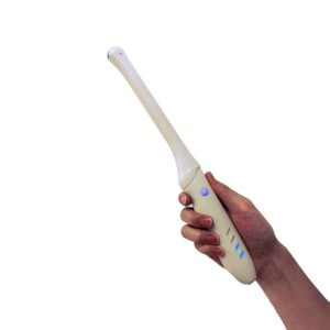
Ultrasound imaging uses high-frequency sound waves to create images of internal organs. Using imaging technology, clinicians are able to detect anomalies and make accurate diagnoses.
Transvaginal ultrasounds, which are also called endovaginal ultrasounds, allow doctors to see inside a woman’s pelvis and uterus. The female reproductive system consists of the uterus, fallopian tubes, ovaries, cervix, and vagina.
The term “transvaginal” refers to an entrance that occurs via the vagina. As the name implies, this is an in-house review.
In this treatment, your doctor or technician will put an ultrasound probe about 2 to 3 inches into your vaginal canal. This is different from a standard abdominal or pelvic ultrasound, in which the ultrasound wand (transducer) is on the outside of the pelvis.
When does one use a transvaginal ultrasound?
The following are some of the situations in which a transvaginal ultrasound would be useful:
- any irregularities in the pelvic or abdominal area
- Possible causes of abnormal pelvic discomfort and bleeding include an ectopic pregnancy (which occurs when the fetus implants outside of the uterus, usually in the fallopian tubes)
- infertility
- verification of correct IUD placement, including screening for uterine fibroids and cysts.
Transvaginal ultrasound is an option your doctor may suggest during pregnancy if you’re having trouble with:
- Keep an eye on the fetus’s heart rate.
- Check the cervix for any signs of change that might indicate an impending miscarriage or early birth.
- look for anomalies in the placenta
- and localize the source of any abnormal bleeding.
- find out if you’re having a miscarriage
- verify a prenatal pregnancy
Exactly what should I do to be ready for a transvaginal ultrasound?
Getting ready for a transvaginal ultrasound is typically not a big deal.
Once you’ve arrived at the clinic or hospital and entered the examination room, you’ll be asked to undress from the waist down and given a gown to wear.
It’s possible that your doctor will tell you to have a partially full or empty bladder for the ultrasound, but this will depend on the reason(s) for the procedure. Intestinal obstructions can be alleviated and pelvic organs can be better seen when the bladder is full.
In order to have a full bladder for your treatment, you should consume around 32 ounces of water or another beverage about an hour beforehand.
Before having an ultrasound, women who are menstruating or who are spotting must remove any tampons they may be using.
What occurs during a transvaginal ultrasound?
At the start of the operation, you will be asked to lie face down on the examination table with your knees bent. When it comes to stirrups, it’s possible but not guaranteed.
The ultrasound wand is lubricated and covered with a condom before being inserted into the vagina by your doctor. Let your doctor know if you have a latex allergy so that they can use a probe cover made out of a material other than latex if one is required.
Pressure might be felt while the transducer is inserted by your doctor. The pressure is comparable to that experienced when a speculum is inserted into the vagina for a Pap smear.
Once the transducer is implanted, it sends images of the pelvic area to a screen through sound waves bouncing off internal organs.
While the transducer is still inside of you, the technician or doctor will slowly rotate it. In doing so, a complete view of your inside organs is obtained.
Sonography through a saline infusion may be ordered by your doctor (SIS). This is a subset of transvaginal ultrasound in which sterile salt water is inserted into the uterus prior to the ultrasound to better see the uterine anatomy.
Slightly stretching the uterus with saline solution allows for a more accurate ultrasound image of the uterine cavity than is possible with standard ultrasound.
Women who are pregnant or have an infection do not qualify for SIS, but a transvaginal ultrasound does.
Where can I find information about the potential dangers of this operation?
Transvaginal ultrasonography is completely safe and has no known adverse effects.
Both the mother and the unborn child are safe during transvaginal ultrasounds. Since this imaging method does not employ radiation, this is the case.
When the transducer is put into the vagina, it will put pressure on the body and, in rare cases, cause discomfort. In most cases, a little discomfort is to be expected, and it should subside after the treatment is through.
Don’t be shy about letting the doctor or technician know if you’re experiencing any severe discomfort.
So, what do we learn from this?
Depending on how quickly your doctor can do the ultrasound, you may get the findings right away. A radiologist reviews the photos taken by a technician during a procedure. Your doctor will receive the report from the radiologist.
Transvaginal ultrasound can be used to find out about a lot of different problems, such as:
- Reproductive system cancer
- routine pregnancies
- cysts \sfibroids
- infected pelvis
- miscarriage due to an unnatural location
- abortion due to placenta previa (a low-lying placenta during pregnancy that may warrant medical intervention)
- Discuss your findings with your healthcare provider to determine the best course of therapy.
Outlook
There are very few hazards involved with a transvaginal ultrasound; nonetheless, you may feel some discomfort. The entire process takes 30–60 minutes, and findings are usually available after 24 hours.
In the event that your doctor is unable to acquire a clear image, you may be asked to return for more testing. Depending on your symptoms, a pelvic or abdominal ultrasound may be performed before a transvaginal ultrasound.
A transabdominal ultrasound may be performed if a transvaginal ultrasound causes you unnecessary pain. For this procedure, your doctor will put gel on your stomach and use a portable instrument to examine your reproductive organs.
-
Product on sale
 Wireless Transvaginal Ultrasound: Vsono-TVU2$2,167.83
$2,614.85 -
Product on sale
 Color Doppler Transvaginal Ultrasound Scanner: Vsono-TVU1$3,016.30
$3,184.48 -
Product on sale
 Double Head Wireless Ultrasound: Vsono-CL3$3,036.92
$3,493.70


